Accessory Navicular Removal
What is Accessory Navicular Removal?
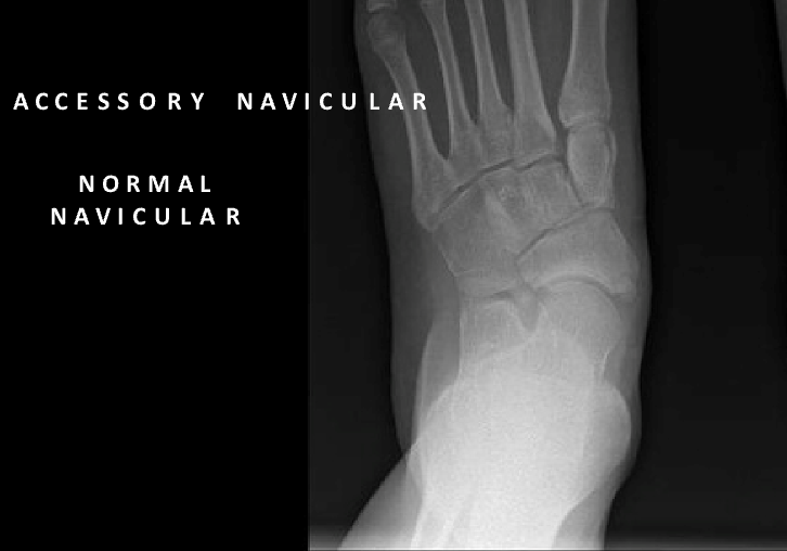
Accessory navicular removal is a surgical procedure designed to address issues related to an accessory navicular, an extra bone or cartilage on the foot's inner side. This congenital condition can cause pain, inflammation, and difficulty with physical activities due to the bone rubbing against shoes or straining nearby tendons. The procedure typically involves removing the accessory bone and, if necessary, repairing or tightening the tibialis posterior tendon that may be affected by the condition.
Who is Suitable for Accessory Navicular Removal?
Accessory navicular removal is recommended for individuals who experience persistent pain, swelling, or discomfort that does not improve with non-surgical treatments. Suitable candidates include:
- Patients with Chronic Pain: Those with ongoing pain that interferes with daily activities or sports despite conservative treatments like orthotics or physical therapy.
- Individuals with Severe Inflammation: Swelling and tenderness around the accessory navicular that does not respond to anti-inflammatory medications or immobilisation.
- Active Individuals: Athletes or physically active people whose performance is hindered by the condition.
- Failed Conservative Treatments: If non-surgical interventions, such as rest, ice, or footwear modifications, fail to alleviate symptoms.
Benefits of Accessory Navicular Removal
- Pain Relief: Eliminating the accessory bone typically resolves pain associated with the condition.
- Improved Mobility: Patients can regain full use of the foot, allowing them to resume normal activities without discomfort.
- Enhanced Foot Function: The surgery can restore proper tendon mechanics, improving the foot’s strength and stability.
- Cosmetic Improvement: For some, removing the protruding bone improves the foot's appearance.
- Long-term Solution: It prevents recurring inflammation and pain by permanently removing the source of irritation.
Types of Accessory Navicular Removal
There are a few surgical techniques that may be used depending on the severity of the condition:
- Kidner Procedure: This is the most common approach. The accessory bone is removed in this procedure, and the tibialis posterior tendon is reattached or repaired to restore function.
- Simple Excision: In this procedure, the accessory navicular is surgically removed without extensive tendon repair. It is often used for less complex cases.
- Osteotomy: If the accessory navicular causes misalignment, the surgeon may perform an osteotomy to realign the bones in the foot.
- Tendon Realignment or Augmentation: If the tibialis posterior tendon is damaged, additional procedures may be performed to repair or strengthen it.
Alternative Options to Accessory Navicular Removal
Surgical removal of the accessory navicular is not the only option. Non-surgical treatments can often manage symptoms effectively, including:
- Orthotics and Arch Supports
- Physical Therapy
- Anti-inflammatory Medications
- Immobilisation
- Corticosteroid Injections
- Lifestyle Modifications
Preparation Before Accessory Navicular Removal
Accessory Navicular removal requires a preoperative medical evaluation. This evaluation consists of a complete patient history and physical examination to determine suitability for the procedure. It may also include lab tests and X-ray imaging.
The surgeon also determines the different surgical procedure options on a case-by-case basis based on the severity of the bursa formation, the patient’s health, and lifestyle.
To determine the best outcome, the individual should be adequately counselled to ensure that the informed consent obtained is fully understood.
The patient is also advised to stop using any anti-inflammatory medications (NSAIDs) at least seven days before the procedure.
Accessory Navicular Removal Procedure
The Accessory Navicular removal surgery is performed under general anesthesia. The steps include:
- Making the individual comfortable in the surgical suite,
- Identification of the proper limb and joint and surgical draping,
- Administration of general anesthesia,
- Surgical incision over the Accessory Navicular bone,
- Complete removal of the Accessory Navicular bone,
- Reattachment of the tibial tendon,
- Closing the incision with stitches and recovery from the anesthesia.
The entire procedure can take two to three hours.
What to Expect After Accessory Navicular Removal?
The Hospital Stay
- You wake up with bulky bandages, a boot
- You will stay in the hospital overnight, with your foot elevated, and you will have antibiotics through a drip.
- Depending on your medical conditions, you will need to take medications to thin your blood for 6 weeks to prevent blood clots (DVT)
- You will need to take vitamin C 1g daily for 6 weeks
- You will be only allowed to touch your foot to the ground for 2-3 weeks
- Depending on your balance and strength, you may need rehabilitation postoperatively
- Buying a second-hand knee scooter pre-operatively (you can search online) and practising at home before the surgery can be helpful; please bring it into the hospital with you. It is easier to use a knee scooter than crutches
Accessory Navicular Removal Rehabilitation
All patients are different. These timelines are only a guide, and some patients may progress faster or slower than others.
- You will need a boot for 6 weeks
- You will have appointments with Dr Graff at 2 weeks and 6 weeks
0 - 3 Weeks
- You will be in a boot
- You can only touch your foot to the ground for balance.
- You will need to bag the leg for showers.
- Pain relief: Please take regular paracetamol with meals and before bed; you may need stronger painkillers as well, especially before bed
- Please take blood thinning medication and vitamin C as prescribed for 6 weeks
2 - 3 Weeks
- Post-op appointment with Dr Graff
- You will then go back into the boot to wear at all times except for showering
- You can start partial weight-bearing in the boot (20-50% body weight)
- You can take the boot off for showers only to sit on a shower chair and keep the foot pointed in
- You can start physio for hip and knee strengthening
6 - 8 Weeks
- Post-op appointment with Dr Graff
- You can weight bear as tolerated in the boot
- You can take the boot off for sleeping
- You will need physiotherapy for active range of motion, proprioception training, and isometric tib post-strengthening
9 - 12 Weeks
- You can wear normal shoes if you can fit into them you (may still have swelling)
- You should wear a supportive lace-up ankle brace and a medial arch support with supportive sneakers
3-6 Months
- Post-op appointment with Dr Graff
- You can progress strengthening and range of motion with pain-free eccentric tib post-strengthening with physiotherapy
- From 4 months, light jogging can commence if there is no pain
- You can walk barefooted
6-12 Months
- When the leg feels normal and the same as the other, you can start sport-specific training and heavy labour work.
When can I return to work/school?
- Seated work 4-6 weeks
- Prolonged standing 10-12 weeks
- Heavy labour work 6 months
When can I return to sport?
- Start sport-specific training at 4-6 months
- Return to sport when the leg is the same as the other side (9-12 months)
When can I drive?
- Left foot 3 weeks (if driving an automatic car)
- Right foot 10-12 weeks
Accessory Navicular Removal Prognosis
Accessory navicular removal generally alleviates symptoms associated with an accessory navicular bone. Patients typically experience significant pain relief and improved foot function post-surgery. Studies indicate high patient satisfaction rates and low complication rates following the procedure.
Accessory Navicular Removal Risks
Accessory Navicular removal carries all the risks associated with major surgery, such as:
- Anaesthetic problems
- Nerve injury
- Blood clots
- Infection
- Stiffness
- Rupture/re-injury
- Ongoing pain
- Further surgery
Delaying Accessory Navicular Removal
Delaying surgical treatment for a symptomatic accessory navicular can lead to ongoing discomfort and may exacerbate associated conditions like posterior tibial tendonitis. Prolonged symptoms can impair daily activities and cause further foot deformities due to altered biomechanics. Early intervention is advisable to prevent these complications.
Contact Us
If you want more information or have any questions or problems, please contact Dr Graff at admin@christygraff.com or call the rooms at 0493 461 133.